Cellomics Core Facility
세포체학실험실

- Facilities
- Cellomics Core Facility
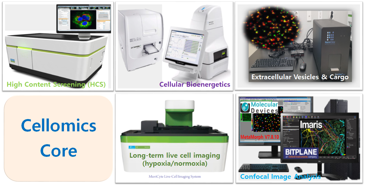
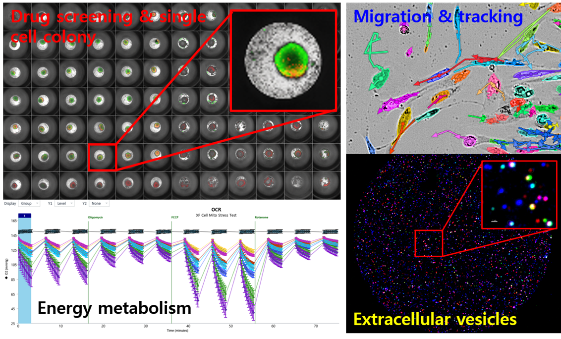
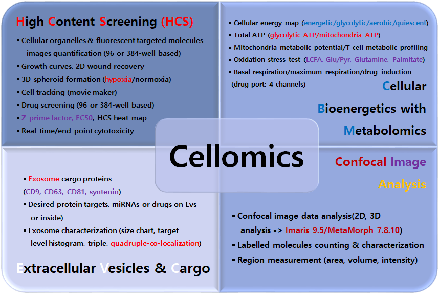
Cellomics is the research discipline of quantitative cell analysis with simultaneous readout of multiple parameters based on phenotypic screening of cells and molecules thereof. CMI Cellomics Core Facility concurrently provides PhD level expertise in technical assistance and scientific support for your research with realistic cell images, objective data analysis and quantitative data visualization acquired by multi-well plate based imaging equipment and extracellular flux analyzer. The service area covered by Cellomics Core Facility is largely divided into four parts:
⊙ Research Equipment:
1. PerkinElmer Operetta CLS (HCS, 4 channels confocal and digital phase contrast)
2. Agilent Seahorse XFe96 analyzer (mitoATP/glycoATP, MitoStress, mito respiration)
3. BioTek Cytation1 (Cell imaging multi-mode reader)
4. Nanoview Biosciences ExoView R100 (exosome tetraspanins, cargo)
5. Revvity MuviCyte (hypoxia/normoxia long-term observation, growth curve)
⊙ Image Analysis Software:
1. Molecular Device MetaMorph V. 7.8.10
2. Oxford Bitplane Imaris V. 9.5
3. PerkinElmer Harmony V. 4.9
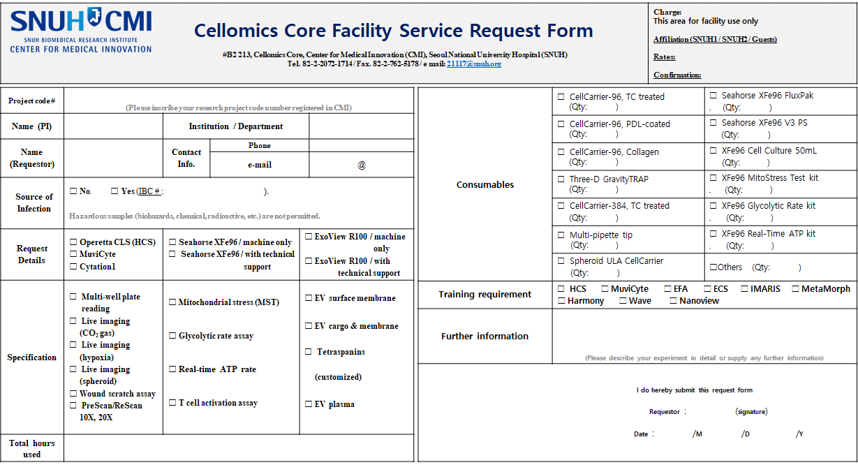
For application of the facility, a service request form should be filled out and submitted to 21117@snuh.org or visit the facility in person. Once your request form is received, further discussion regarding time schedule, methods, reports and fees will be made by discussion with lab manager. If there is a need for further discussion with reference to the HCS or XFe-96 experimental design, contact us by phone (ext. 1714) or visit the facility with reference papers.
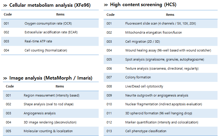
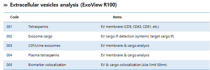
⊙ Items for analysis:
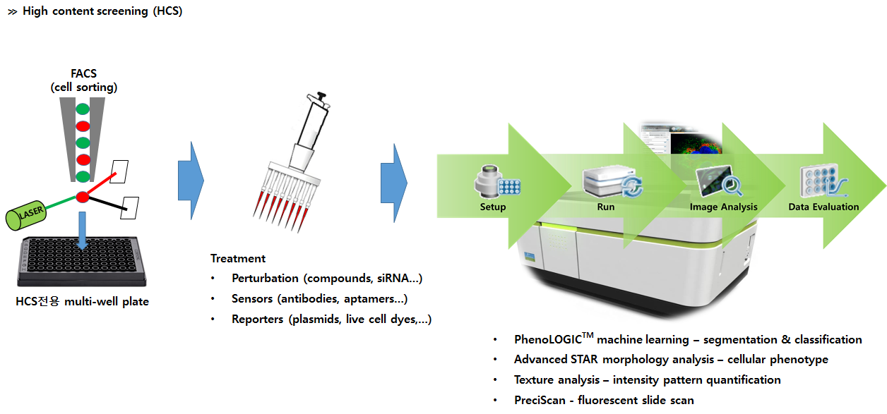
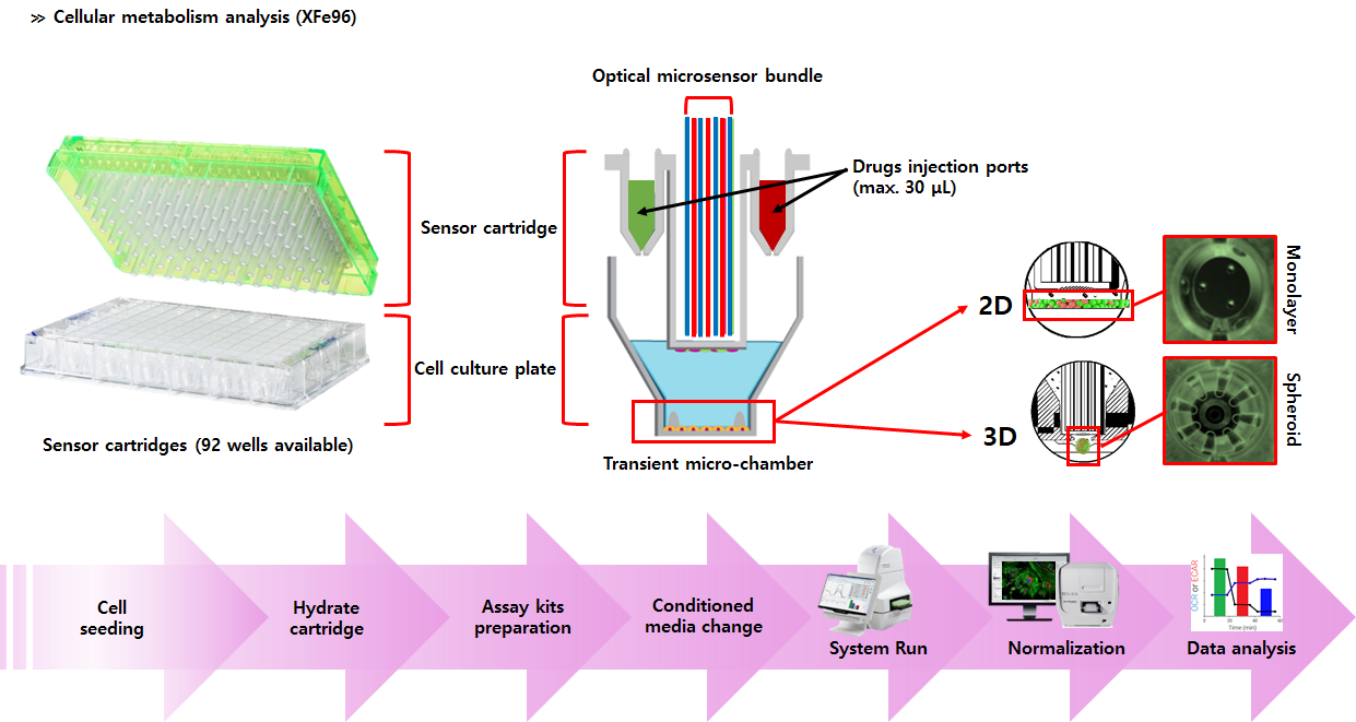
⊙ Processing
Mailing address and for questions and information:
For Operetta CLS (HCS), extracellular flux analyzer (Seahorse Xfe-96), and image analysis inquiries:


